
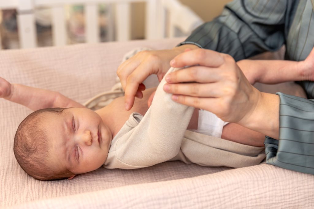
Obstetric brachial plexus palsy can initially be frightening for parents. The newborn’s arm remains still alongside the body, held in internal rotation with the elbow extended and the wrist and fingers flexed. Sometimes, sensory disturbances may also be present in the affected upper limb. Recovery rates range from 80% to 96%, particularly when treatment begins within the first two weeks of the child’s life.
Obstetric brachial plexus palsy is divided into two main types according to the distribution and severity of the injury. Upper-type paralysis (Erb’s palsy) involves the C5 and C6 nerve roots and, in some cases, C7 as well. Total obstetric brachial plexus palsy involves the nerve roots from C5 to T1. Klumpke’s palsy affects the C8 and T1 nerve roots.
The injury involves stretching of the nerves of the brachial plexus due to a force of separation or traction between the head and shoulder. Causes of obstetric brachial plexus palsy may include:
- Shoulder dystocia
- Macrosomia (birth weight over 4 kg)
- Fetopelvic disproportion, with a narrow maternal pelvis and/or maternal overweight or obesity (associated with diabetes mellitus or gestational diabetes)
- Breech delivery
Treatment may be conservative and may include physiotherapy (Neurodevelopmental Treatment, such as Bobath and Vojta, electrical stimulation, and joint mobilization techniques), alternative or holistic therapies (e.g., Ayurveda), botulinum toxin (Botox) injections, or surgery. Surgery is generally considered in cases where there has been no significant improvement by nine months of age. In severe cases, surgical treatment may be recommended as early as two months of age.
As the child develops, the muscle imbalance between the agonist and antagonist muscles of the upper limb affects not only movement but can also lead to changes in the bones and joints. The most common abnormalities affect the glenohumeral joint (shoulder joint) and the shoulder girdle (clavicle and scapula). In upper-type paralysis, the affected external rotators of the humerus are unable to counterbalance the action of the internal rotators. This results in anterior displacement of the humeral head and internal rotation of the arm, leading to adaptive changes in the glenohumeral joint. Changes may also occur at the elbow due to weakness of forearm supination and full elbow extension, as a result of paralysis of the corresponding muscles, as well as at the metacarpophalangeal joints of the fingers.
As mentioned above, treatment should ideally begin during the first month of life. But which treatment is most appropriate? Some studies have reported that conventional treatments recommended for children, such as the Bobath and Vojta approaches or forced use of the affected limb, do not produce particularly significant results. The use of electrical stimulation remains controversial, although some studies support its effectiveness.
Because obstetric brachial plexus palsy affects the musculoskeletal system and can result in adaptations and changes such as muscle shortening, reduced joint range of motion, joint instability, and muscle contractures, it is understandable that treatments involving joint mobilization, muscle stretching, fascial release, and reduction of muscle spasms may produce more immediate benefits.
To understand why, it is useful to consider what happens when we raise our arm overhead to 180°. Simultaneous movements occur at several joints and interfaces: • The joint between the clavicle and sternum (sternoclavicular joint) • The joint between the clavicle and scapula (acromioclavicular joint) • The interface between the scapula and thorax (scapulothoracic articulation) • The glenohumeral joint. The 180° of upper-limb abduction consists approximately of 120° of glenohumeral movement and 60° of scapular upward rotation. When we initially begin raising our arm, the scapula moves very little and is relatively stabilized against the thorax. This occurs during approximately the first 60° of shoulder flexion and the first 30° of abduction. Corresponding changes also occur at the other joints mentioned above. These movements occur in a coordinated relationship known as the scapulohumeral rhythm, which is controlled by various structures both within and around the shoulder joint.
In obstetric brachial plexus palsy, the scapulohumeral rhythm can be significantly affected. The muscles may be unable to stabilize the scapula properly against the thorax, causing the scapula to move abnormally as the child raises their arm. When the arm does not move through its full range of motion, the structures both inside and outside the joint may gradually shorten and permanently restrict movement. In these cases, joint mobilization and relaxation of the surrounding soft tissues can be particularly important. Joint mobilization techniques and soft-tissue techniques are widely used in the treatment of adults. But what about treating children, and particularly infants?
What is most important for the appropriate management of obstetric brachial plexus palsy is that the therapist has a thorough understanding of the function of the joints described above, the pathology that can result from impaired joint function, and the imbalance that may develop throughout the body if the condition is not addressed appropriately and in a timely manner. Only with this understanding can the therapist design an appropriate treatment plan. Combining knowledge of approaches such as Bobath, which are used with infants and children, with techniques applied in manual therapy, may help achieve the best possible outcomes. The use of rigid tape or kinesiology tape (kinesiotape) may also help maintain treatment outcomes. All of these interventions should be incorporated into a play-based environment, giving the child opportunities to use the abilities gained through physiotherapy in everyday activities.
References:
Bahm, J., Ocampo-Pavez, C., Disselhorst-Klug, C., Sellhaus, B. & Weis J. (2009). Obstetric Brachial Plexus Palsy Treatment Strategy, Long-Term Results, and Prognosis. Deutsches Ärzteblatt International, 106(6), 83–90.
Chater, Μ., Camfield, P. & Camfield C. (2004). Erb’s palsy – Who is to blame and what will happen? Paediatric Child Health, 9(8), 556-560.
Okafor, U.A., Akinbo, S.R., Sokunbi, O.G., Okanlawon, A.O. & Noronha, C.C. (2008). Comparison of electrical stimulation and conventional physiotherapy in functional rehabilitation in Erb’s palsy. Nigerian Quarterly journal of hospital medicine, 18(4):202-5.
Srilakshmi, D. & Chaganti, S. (2013) A holistic approach to the management of Erb’s palsy. Journal of Ayurveda & Integrative Medicine, 4 (4).